Friday, April 29, 2016

Fungi Observations Group Post by: Taylor Jones, Amy Latimer, Lisa Merritt, Shawna Steele


The mushroom our group cultivated was the Lion's Mane mushroom (Hericium erinaceus), which most in our group had never heard of so it was interesting to look up facts about this mushroom. Lion's mane of course is in  
Kingdom Fungi,
Division Basidiomycota
Subdivision Agaricomycetes--Nearly all species are terrestrial (a few are aquatic), occurring in a wide range of environments where most function as decayers, especially of wood. However, some species are pathogenic or parasitic, and yet others are symbiotic (i.e., mutualistic), these including the important ectomycorrhizal symbionts of forest trees.
Order: Russulales which represent an independent evolutionary line of agarics, not directly related to the Agaricales. The fungal order Agaricales also known as gilled mushrooms, which are known for their distinctive gills or euagarics. Agaricales contains some of the most familiar types of mushrooms.
Genus: Hericium which are known for their medicinal properties in oriental medicine and  fruiting bodies with pegs, spines or teeth hanging from their hymenium. Lion's Mane can be identified by its long spines (greater than 1 cm length), its appearance on hardwoods and its tendency to grow a single clump of dangling spines.
Lion's mane is native to North America, Europe and Asia  Lion’s Mane fruiting begins within 1-2 weeks of initiating the kit. Blocks will produce mushrooms over the course of 2-4 flushes for 1-2 months, depending on care.


The Mushroom kit was set up during lab one. Where the kit was set up according to the instructions. Observations were taken on April 20th, 25th, and 27th. As you can see the Lion’s Mane made some progress in growth every week with the most growth observed on the 27th.
On week one of observations a small amount of growth was observed where floret-esque bodies were starting to form.
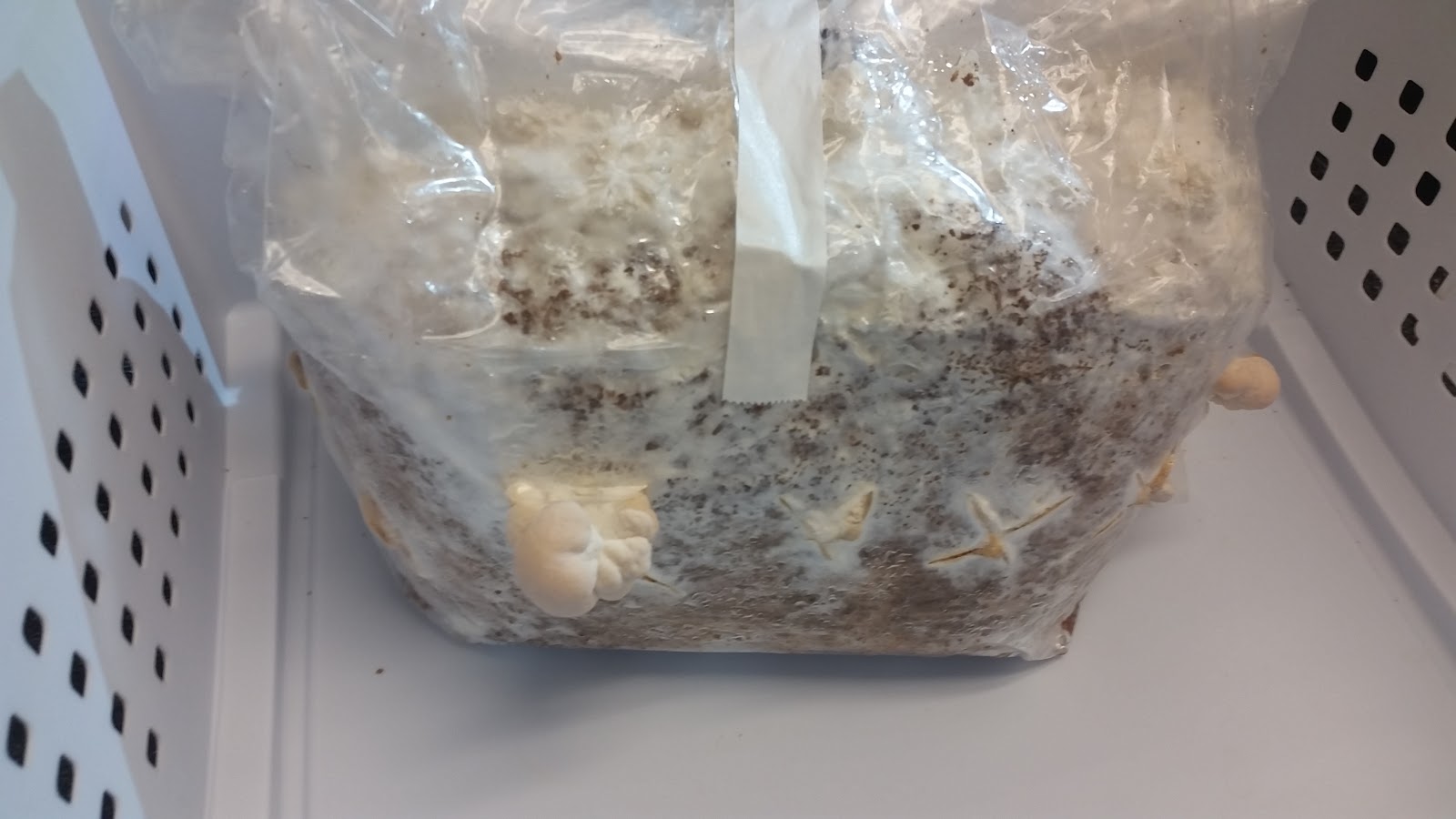
Image 1: Lion’s Mane observation April 20, 2016
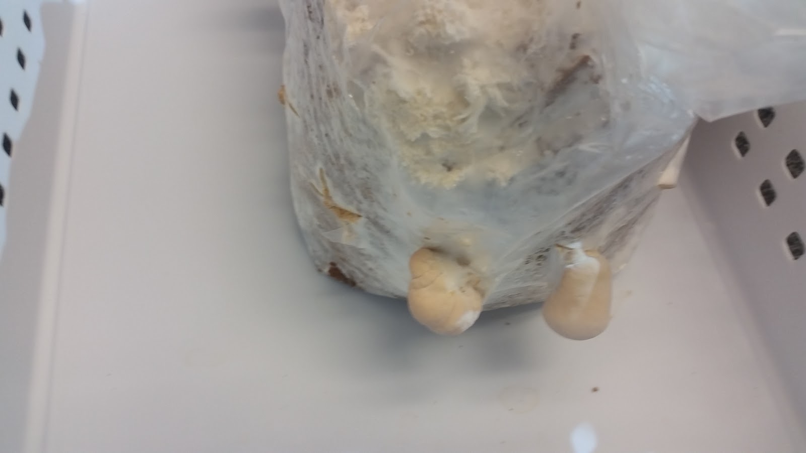
          Image 2: Lion’s Mane observation April 20, 2016

Image 3:  Lion’s Mane observation April 20, 2016


On April 25th The floret-esque bodies became bigger in size and additional bodies were starting to grow!
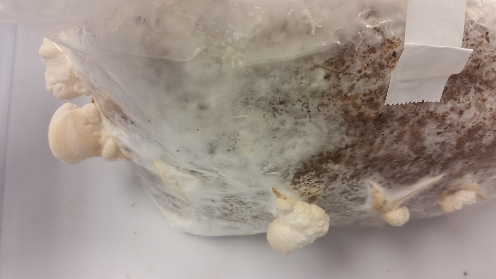
Image 4: Lion’s Mane observation April 25, 2016
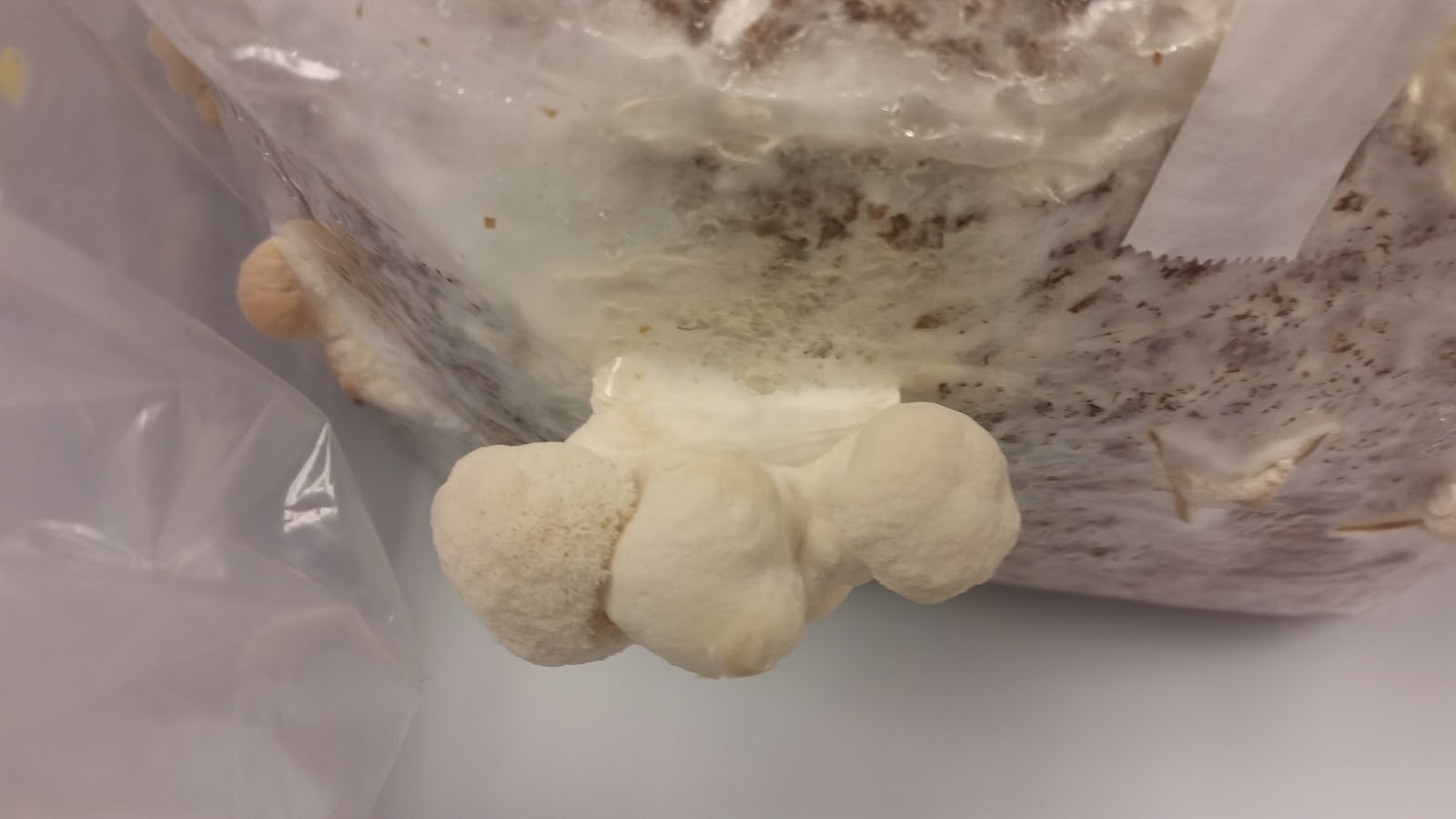
Image 5: Lion’s Mane observation April 25, 2016.
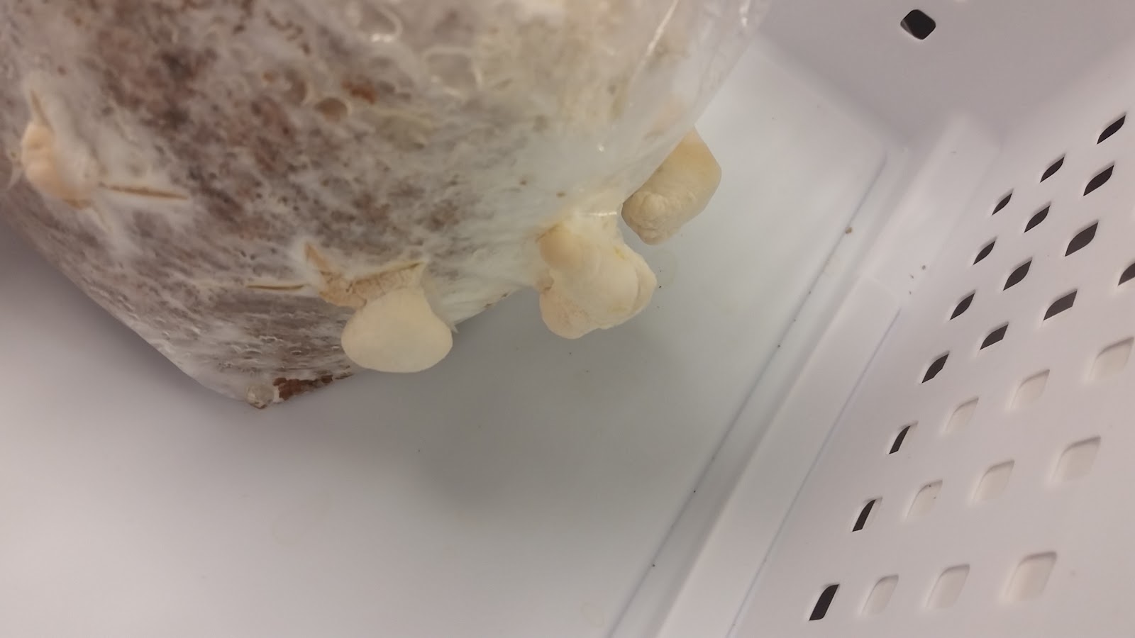
Image 6: Lion’s Mane observation April 25, 2016.

The last observation of the Lion’s Mane was taken on April 27th.  The already growing florets grew bigger in size. Also, as seen in image 9 new bodies started to grow. In the weeks we observed the Lion’s Mane steady growth and new body formation was observed.
Image 7: Lion’s Mane observation April 27, 2016
       
                                                  Image 8: Lion’s Mane observation April 27, 2016


Image 9: Lion’s Mane observation April 27, 2016


IMG_2762.JPG
Image 10. Lion’s Mane observation April 27, 2016


Unfortunately, we couldn’t prepare a thin enough slide to examine our Lion’s Mane under a microscope.  Image 11 was the best we could do. As you can see, there is really nothing to see except the shadow outline of the piece we sliced up. Oops.
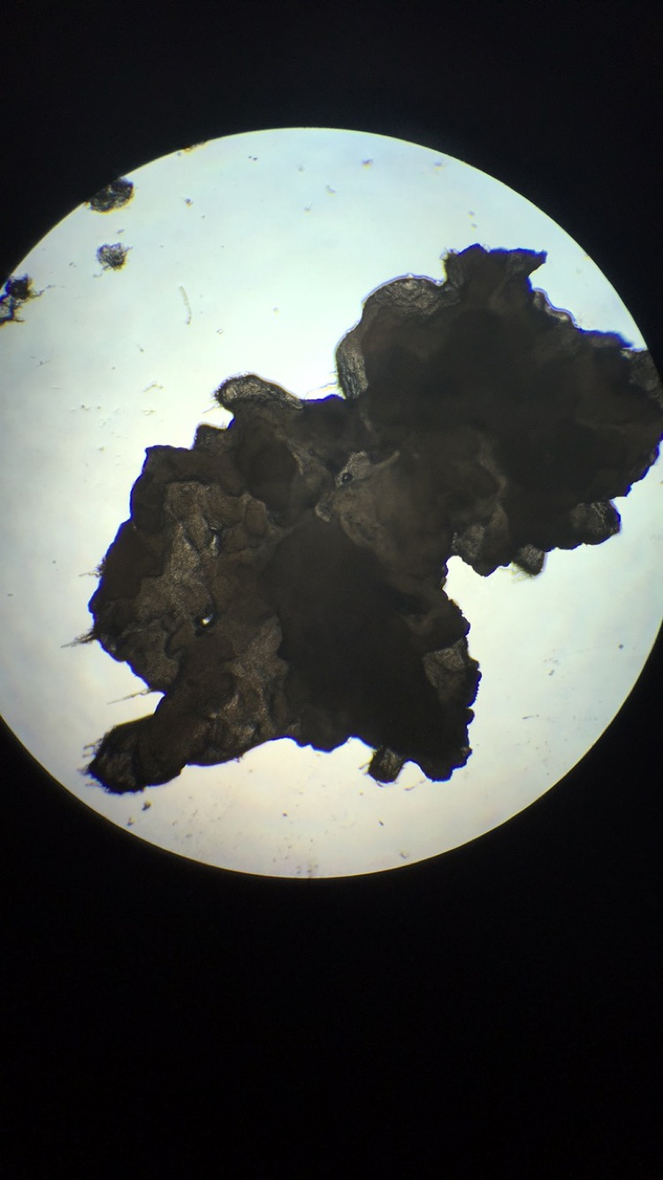

Image 11. Lion’s Mane observation April 27, 2016 - failed attempt

Pilobolus also know as Pilobolus crystallinus. Of course also belongs to
Kingdom: Fungi unlike the Lion’s Mane Pilobolus belongs to the division of Zygomycota. Like the Lion’s mane
Zygomycota are mostly terrestrial in habitat, however, Zygomycota  live in soil or on decaying plant or animal material. Some are parasites of plants, insects, and small animals, while others form symbiotic relationships with plants.
Class: Mucoromycotina which includes many rapidly growing species. Most being saprobes, plant pathogens, and mycoparasites. Some of which induce human mycosis.
Genus: Pilobolus
The life cycle of Pilobolus begins with a black sporangium that has been discharged onto a plant such as grass. A herbivorous animal such as a cow  then eats the grass and unknowingly consumes the sporangium The Pilobolus sporangium passes through the gastrointestinal tract without germinating, and surfaces with the excrement. Once outside its host, spores within the sporangium germinate and grow as a mycelium within the excrement, where it performs as a colonizer.. Later, the fungus fruits to produce more spores. The asexual fruiting structure (the sporangiophore) of Pilobolus is unique in that . It consists of a transparent stalk which rises above the excrement to end in a balloon-like sub sporangial vesicle on top of this, a single, black sporangium that develops. The sporangiophore orients  itself to point directly towards a light source. The sub sporangial vesicle acts as a lens that focuses light via carotenoid pigments deposited near the base of the vesicle. The developing sporangiophore grows such that the maturing sporangium is aimed directly at the light.
When turgor pressure within the sub sporangial vesicle is at an ample amount the sporangium is launched, and can travel anywhere from a couple of centimeters to a distance of 2 meters. The details of the Zygomycota  life cycle can be seen in figure 11.
In contrast, the lifecycle of basidiomycetes includes alternation of generations. Spores are produced through sexual reproduction, rather than asexual reproduction. The club-shaped basidium carries spores called basidiospores. In the basidium, nuclei of two different mating types fuse called karyogamy, producing  a diploid zygote that then undergoes meiosis. The haploid nuclei migrate into basidiospores, which germinate and generate monokaryotic hyphae. The resulting mycelium is called a primary mycelium. Mycelia of different mating types can combine and produce a secondary mycelium that contains haploid nuclei of two different mating type this is known as the dikaryotic stage of the basidiomycetes life cycle and it is the dominant stage. After some time, the secondary mycelium generates a basidiocarp, which is a fruiting body that juts from the ground typically what we think of as a  mushroom. The basidiocarp bears the developing basidia on the gills under its cap. The details of the Basidiomycota life cycle are detailed in image 10.


Image 10: Depiction of the life cycle of Basidiomycota
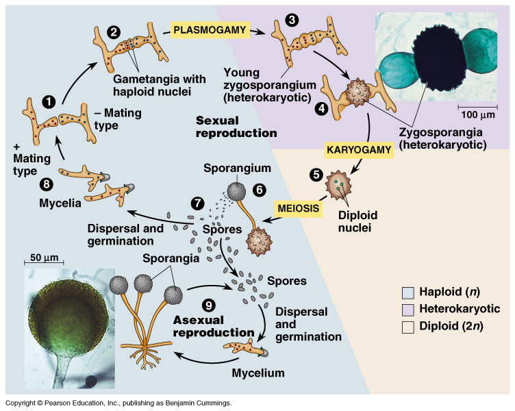
Image 11: Depiction of life cycle of Zygomycota.

IMG_2571.JPG
Image 12: Pilobolus Day 1- Before experiment setup
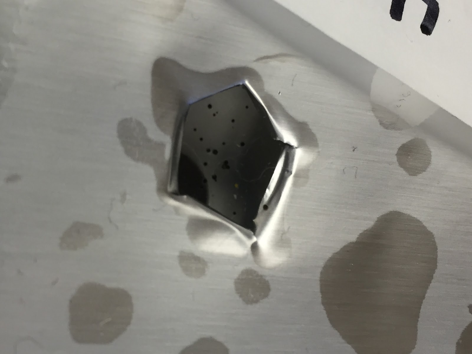

IMG_2667 (1).JPG
Image 13: The result of the experiment, Pilobolus heads that shot towards the light via phototropism

IMG_2686.JPG
Image 14: Pilobolus after growth- experiment results

IMG_2687.JPG
Image 15. Close up of the Pilobolus
IMG_2689.JPG

Image 16. An even closer look of the Pilobolus- experiment results


Fungi Field Trip Post
Tony Tilt


A small Bracket mushroom

 On our field trip we saw many different types of fungi and mushrooms. This one that i have a picture of is a type of bracket mushroom. It was estimated to be about 2 years old and it is a very hard and tough type of mushroom. It has poly pores and it has tiny pore tubes that hold spores. The mushroom decomposes the log and can grow as long as the log is still there.
A larger group of Bracket mushrooms
This is another picture of the same mushroom. These ones were estimated to be about 10 years old. These are also vary hard and you can tap on them and hear how hard they are. These ones will continue growing until the log is totally decomposed and then the mushrooms will just die.

Fungi Culture Observation - From Shiitake to Pilobolus by the Dream Team

Fungal Culture Observation Post by the Dream Team

Group members: Matthew Heinekin, Nhy Tran, Ivanna de Anda

       Mushroom Log Observation:


Shiitake Scientific Classification:
Kingdom: Fungi
Division: Basidiomycota 
Class: Agaricomycetes
Order: Agaricales
Family: Marasmiaceae
Genus: Lentinula
Species: L. edodes

Figure 1. Shiitake mushroom log on day 1

Our group chose a Shiitake mushroom log to grow and expected to see a lot of mushroom would grow. To create a humid environment for the mushroom, we covered the log with a plastic bag and often sprayed water to keep it moist (Figure 1)

Figure 2. Shiitake mushroom log on the final day

Several weeks passed, we didn't see any signs of mushrooms on our log, but we could only see the mushroom log just got browner and browner. Our group's members think that there weren't any mushroom grew because the log was overwatered, and we could see that the surface of the log looked extremely moist and soggy (Figure 2). Or maybe, it didn't want to grow any mushrooms because they don't want to be eaten, who knows!!!

Figure 3. Oyster mushroom gills under microscope (400X)

Since our mushroom log didn't grow any mushrooms, we asked other groups for the mushroom gills and made a wet mount of it (Figure 3). The mushroom that we did was oyster mushroom 

      Pilobolus Observation:


Pilobolus Scientific Classification: 
Domain: Eukaryote
Kingdom: Fungi
Division: Zygomycota
Class: Mucoromycotina
Order: Mucorales
Family: Pilobolaceae 
Genus: Pilobolus 

Figure 4. The growth of Pilobolus after one week

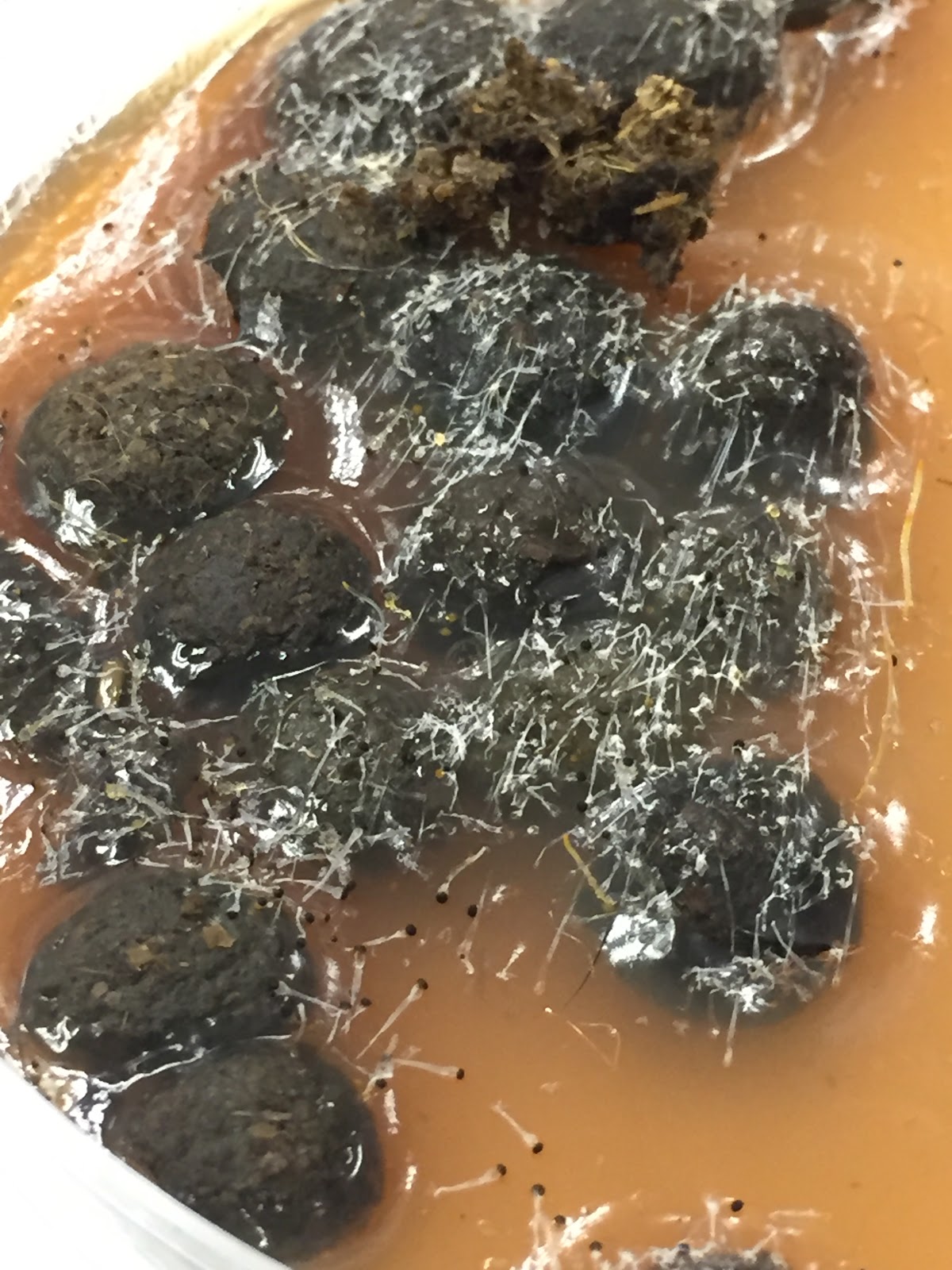
The Pilobolus fungi culture was grown on a fecal petri dish inside a small cup, and it was covered with aluminum foil that had a hole in the center for the light source. This fungi shot its sporangia toward the light (a hole on aluminum foil) and made the cup filled with black dots, which are the spores. 
Figure 5. A close up look of Pilobolus 

     Similarities and differences between Shiitake mushroom and Pilobolus fungi:
- Shiitake is in the division Basidiomycota, which form specialized club-like cells called basidia; but Pilobolus is in the division Zygomycota, which the haploid hyphae from two different individuals met and fuse to form a spore-producing structure called a zygosporangium 
- The mushroom is a lot bigger than the Pilobolus fungi
- Shiitake grows on the log, but Pilobolus grows on the dung (fecal petri dish used in the experiment)
- Shiitake had to be taken care of more than Pilobolus
- Pilobolus discharges its sporangia towards the light source, Shiitake mushroom doesn't do that


Thursday, April 28, 2016

Fungal Culture Observation Post by Group [4.0 A]


Group members: John Yang, Ali Ahmadi, Tony Tilt, Nobnaret Sonthirak (Kyle) 

 Figure 1. illustrates oyster mushroom log on the first day.

Our group did the oyster mushroom. We first sprayed it down with the de-ionized water, and then we covered it with a plastic bag. This is to maintain the humid environment that will maximize the growth of the mushroom. We repeated this for every lab session. 

Initially, there was no fruiting body present on the log. 

Figure 2. displays oyster mushroom log on the final day.
With regards to the final day, there were more mushroom than we could count. The size vary from the biggest being 7 cm to the smallest being 1 cm. Moreover, the bottom of the tray was covered with spores.

Pilobolus Observation

Figure 3. shows the overview of the pilobolus at an early of their development

Figure 4. illustrates the zoom-in version of the pilobolus under a dissecting microscope
Figure 5. shows the spores from the explosion toward the light
 Compare and contrast between oyster mushroom and pilobolus fungi 


- Both of the mushroom grow toward the light (i.e. an of phototropism).
-They secrete spores in different ways. One of them which is oyster mushroom, dropped it's spores onto the tray. There were a lot a white spores on the tray. The pilobolus fungi exploded and releasing black spores to the surrounding environment towards the sunlight. 
-The pilobolus fungi is much smaller than the oyster mushroom. 
-The oyster mushroom produced spores continuously meanwhile the pilobolus fungi produce spores only when it explodes.  


Wednesday, April 27, 2016

Fungi Culture - From Shiitake to Pilobolus by Gwen


The objective of the fungi culture post is to make observation notes of the two different fungi cultures that you took care of and observed in lab.
Here are a few things to focus on in your post:

  • what are the two plylum that you observed?
  • compare and contrast the two species of fungi
  • compare life cycles - similarities and differences
  • how successful were the cultures and why do you think they did/didn't grow well?
  • what does that show you about the ecology of these different fungi?
  • what are some cool things that you observed?
  • what are some unexpected things that you observed?
  • did you think this was a useful assignment? 
    • was it fun? 
    • did you learn something?
    • would you recommend it for next year's students?

Thursday, April 21, 2016

TSA Microorganisms

Day 0:

24 degrees Celsius                                                               37 degrees Celsius


I decided to take swabs from a classroom window, under the desk, under my nail, and between my toes. I suspected that I would find a lot of bacteria, likely good and bad, in the colonies that I expected would grow from all four samples. More colonies were predicted to be found in the 37 degree environment as it is significantly warmer than room temperature.

Day 2:

24 degrees Celsius                                                               37 degrees Celsius


On Day 2, I found only a few colonies in the "under nail" and "between toes" samples in the 24 degree environment and more than 100 colonies each in the same samples placed in the 37 degree environment. This was surprising to me because I expected there to be at least some sort of growth in the "under desk" and "window" samples but no colonies grew.


Day 7:

24 degrees Celsius                                                                 37 degrees Celsius


On the final day, before throwing the TSA plates out, I observed even more colony growth, in size and number of colonies, again mostly on the "under nail" and "between toes" samples. There was about as many colonies in the 24 degree plate as there had been in the 37 degree plate the week before. In the 37 degree plate, colonies grew mainly in size. One large colony grew in the "under desk" sample in the 37 degree plate, and in the same plate grew a very small colony in the "window" sample. Overall, I found myself both disgusted and impressed by the amount of bacteria that grew from between my toes and under my nails and likewise, disappointed by the lack of bacteria that grew from the other samples. I suspect that the lack of growth was due to the fact that the windows and desks are wiped down with antibacterial wipes and cleaning products regularly. All of the colonies that grew appeared to be a light beige/yellow color and were fairly flat against the gel. 

Monday, April 18, 2016

Mystery Bacteria on TSA agar plates by Robert Barker

Day 0
            I started out wondering what organisms were on my wheelchair. I like keeping myself healthy so, I also decided to make sure my tattoo on my shoulder was not covered with unnecessary bacteria. I also took a sample from my tongue. The last item I took a sample of was the telephone in the lab. I made two plates; each of them contained the same samples. One of the plates was left out in room temperature, whereas; the other sample was put into an incubator of 37̊̊ Celsius (body temperature). I hypothesized that the organisms on my wheelchair  would be of great abundance in the body temperature incubator. I  thought that if you took the organisms out of their normal habitat, room temp. or body temp., that they would speed up production. I unfortunately did not take a picture the first day.


Day 2

Figure 1: Plates growth after 2 days of incubation. The left plate was placed at 37 degrees Celsius and the right was in room temperature incubation.

            I entered class and checked up on how the bacteria was growing. I was surprised at the results I obtained. My hypothesis was beginning to be supported but, not as quickly as I thought it would be. My tongue was the only item not supported in my hypothesis. After two days there was no growth on my room temperature plate that I could see. The plate incubated at body temperature had some growth but, the room temperature one did not.

Colonies on body temperature:
Tongue - 2
Wheelchair tire  ̴ 16
Tattoo on shoulder  ̴̴  13
Phone in lab  ̴ 1

Day 7


Figure 2: Plates growth after 7 days. The plate on the left is TSA agar plate that was incubated at body temperature. The plate on the right is incubated at room temperature.

       My hypothesis on why no bacteria grew on the room temperature samples of my tongue, shoulder and on the school phone is because I may have not actually swab the surface before rubbing the q-tip on that plate. 

Colonies on TSA agar Plates

         Room temperature plate                                    Body temperature plate
                                                                                         Tongue  ̴ 4
            Wheelchair is approximately 600 colonies                                                                                                                                                                        Wheelchair Tire  ̴  29 
                                                                                           (based on Figure three)
                                                                                            Tattoo on Shoulder  ̴ 16
                                                                                             Phone in lab  ̴  6 

Closer Image of Wheelchair tire
Figure 3: Image of my wheelchair tire under the dissecting microscope after 7 days of Body temperature environment.

 I was intrigued by all the growth on the wheelchair tire incubated at body temperature. The organism at bottom right, the whitish grey one, looked like cauliflower to me. When looking at the Medical Microbe Gallery 1, I believe I can conclude that the organism is a Nocardia asteroides morphology. I also see Staphylococcus aureus morphology.

Reference used:
1. http://www.microbelibrary.org/component/resource/laboratory-test/3114-colony-morphology?limit=0&limitstart=0