The mushroom our group cultivated was the Lion's Mane mushroom (Hericium erinaceus), which most in our group had never heard of so it was interesting to look up facts about this mushroom. Lion's mane of course is in
Kingdom Fungi,
Division Basidiomycota
Subdivision Agaricomycetes--Nearly all species are terrestrial (a few are aquatic), occurring in a wide range of environments where most function as decayers, especially of wood. However, some species are pathogenic or parasitic, and yet others are symbiotic (i.e., mutualistic), these including the important ectomycorrhizal symbionts of forest trees.
Order: Russulales which represent an independent evolutionary line of agarics, not directly related to the Agaricales. The fungal order Agaricales also known as gilled mushrooms, which are known for their distinctive gills or euagarics. Agaricales contains some of the most familiar types of mushrooms.
Genus: Hericium which are known for their medicinal properties in oriental medicine and fruiting bodies with pegs, spines or teeth hanging from their hymenium. Lion's Mane can be identified by its long spines (greater than 1 cm length), its appearance on hardwoods and its tendency to grow a single clump of dangling spines.
Lion's mane is native to North America, Europe and Asia Lion’s Mane fruiting begins within 1-2 weeks of initiating the kit. Blocks will produce mushrooms over the course of 2-4 flushes for 1-2 months, depending on care.
The Mushroom kit was set up during lab one. Where the kit was set up according to the instructions. Observations were taken on April 20th, 25th, and 27th. As you can see the Lion’s Mane made some progress in growth every week with the most growth observed on the 27th.
On week one of observations a small amount of growth was observed where floret-esque bodies were starting to form.
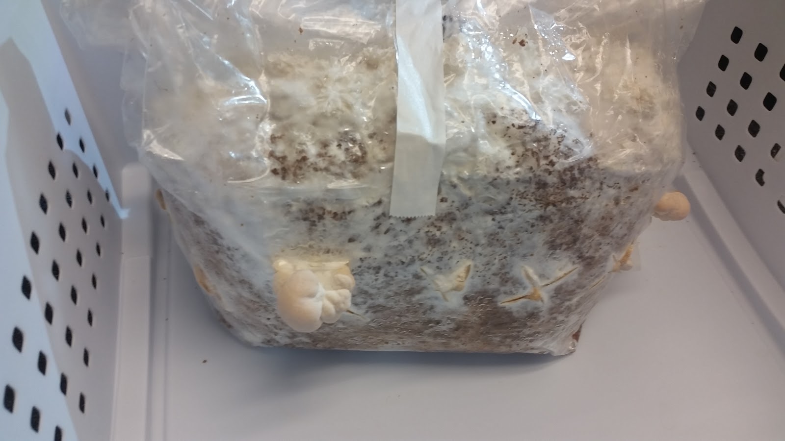
Image 1: Lion’s Mane observation April 20, 2016
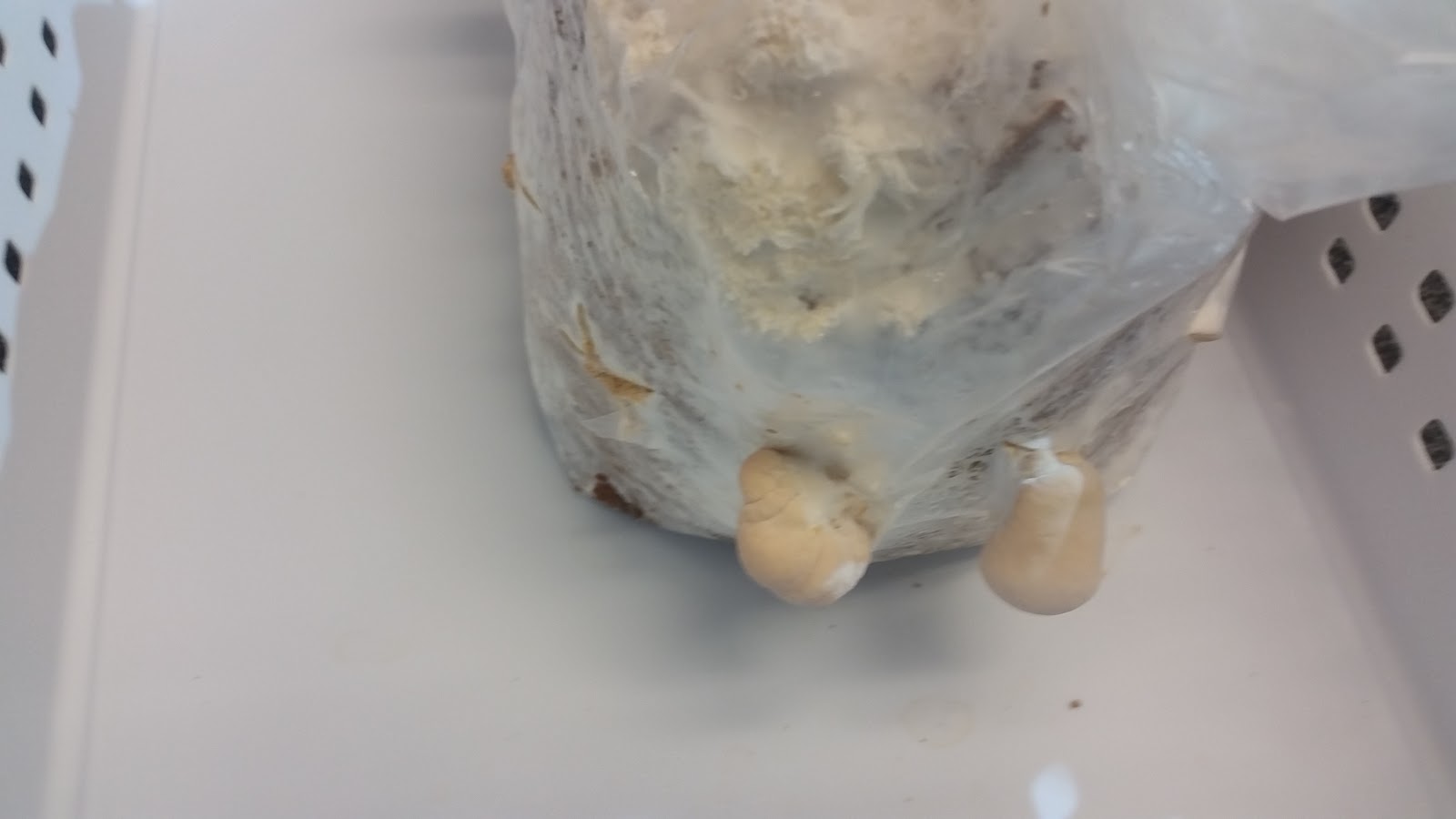
Image 2: Lion’s Mane observation April 20, 2016

Image 3: Lion’s Mane observation April 20, 2016
On April 25th The floret-esque bodies became bigger in size and additional bodies were starting to grow!
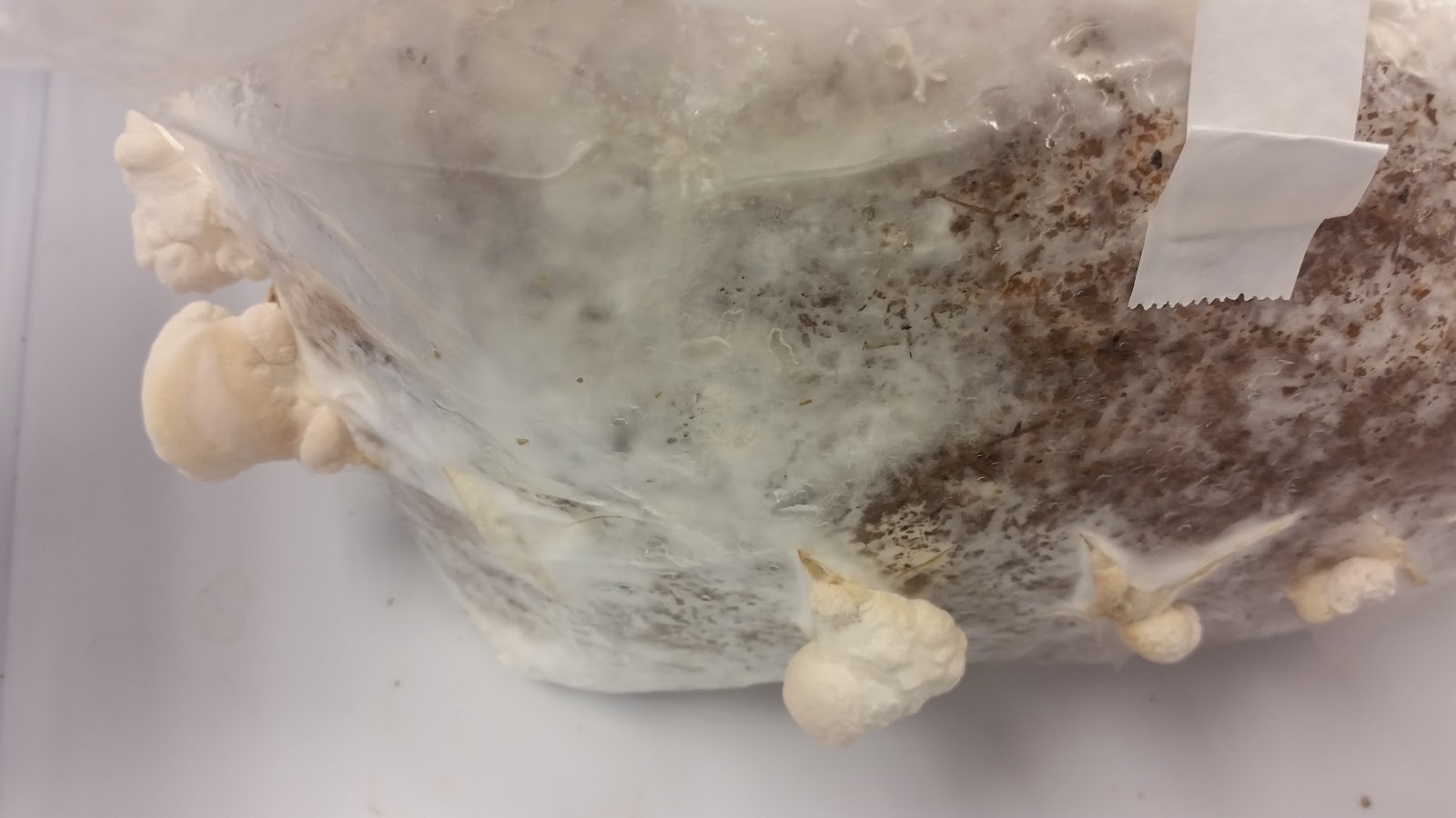
Image 4: Lion’s Mane observation April 25, 2016
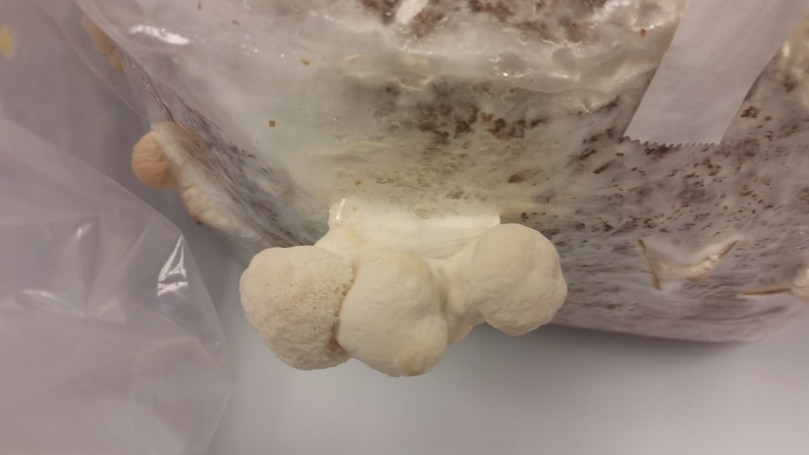
Image 5: Lion’s Mane observation April 25, 2016.
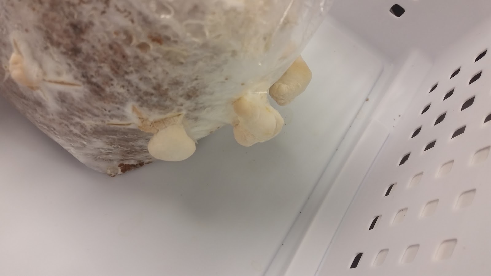
Image 6: Lion’s Mane observation April 25, 2016.
The last observation of the Lion’s Mane was taken on April 27th. The already growing florets grew bigger in size. Also, as seen in image 9 new bodies started to grow. In the weeks we observed the Lion’s Mane steady growth and new body formation was observed.

Image 7: Lion’s Mane observation April 27, 2016

Image 8: Lion’s Mane observation April 27, 2016

Image 9: Lion’s Mane observation April 27, 2016

Image 10. Lion’s Mane observation April 27, 2016
Unfortunately, we couldn’t prepare a thin enough slide to examine our Lion’s Mane under a microscope. Image 11 was the best we could do. As you can see, there is really nothing to see except the shadow outline of the piece we sliced up. Oops.
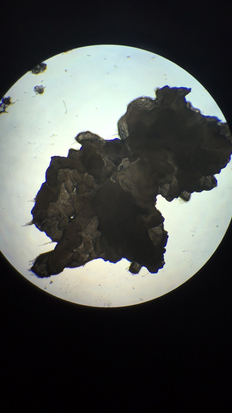
Image 11. Lion’s Mane observation April 27, 2016 - failed attempt
Pilobolus also know as Pilobolus crystallinus. Of course also belongs to
Kingdom: Fungi unlike the Lion’s Mane Pilobolus belongs to the division of Zygomycota. Like the Lion’s mane
Zygomycota are mostly terrestrial in habitat, however, Zygomycota live in soil or on decaying plant or animal material. Some are parasites of plants, insects, and small animals, while others form symbiotic relationships with plants.
Class: Mucoromycotina which includes many rapidly growing species. Most being saprobes, plant pathogens, and mycoparasites. Some of which induce human mycosis.
Genus: Pilobolus
The life cycle of Pilobolus begins with a black sporangium that has been discharged onto a plant such as grass. A herbivorous animal such as a cow then eats the grass and unknowingly consumes the sporangium The Pilobolus sporangium passes through the gastrointestinal tract without germinating, and surfaces with the excrement. Once outside its host, spores within the sporangium germinate and grow as a mycelium within the excrement, where it performs as a colonizer.. Later, the fungus fruits to produce more spores. The asexual fruiting structure (the sporangiophore) of Pilobolus is unique in that . It consists of a transparent stalk which rises above the excrement to end in a balloon-like sub sporangial vesicle on top of this, a single, black sporangium that develops. The sporangiophore orients itself to point directly towards a light source. The sub sporangial vesicle acts as a lens that focuses light via carotenoid pigments deposited near the base of the vesicle. The developing sporangiophore grows such that the maturing sporangium is aimed directly at the light.
When turgor pressure within the sub sporangial vesicle is at an ample amount the sporangium is launched, and can travel anywhere from a couple of centimeters to a distance of 2 meters. The details of the Zygomycota life cycle can be seen in figure 11.
In contrast, the lifecycle of basidiomycetes includes alternation of generations. Spores are produced through sexual reproduction, rather than asexual reproduction. The club-shaped basidium carries spores called basidiospores. In the basidium, nuclei of two different mating types fuse called karyogamy, producing a diploid zygote that then undergoes meiosis. The haploid nuclei migrate into basidiospores, which germinate and generate monokaryotic hyphae. The resulting mycelium is called a primary mycelium. Mycelia of different mating types can combine and produce a secondary mycelium that contains haploid nuclei of two different mating type this is known as the dikaryotic stage of the basidiomycetes life cycle and it is the dominant stage. After some time, the secondary mycelium generates a basidiocarp, which is a fruiting body that juts from the ground typically what we think of as a mushroom. The basidiocarp bears the developing basidia on the gills under its cap. The details of the Basidiomycota life cycle are detailed in image 10.

Image 10: Depiction of the life cycle of Basidiomycota
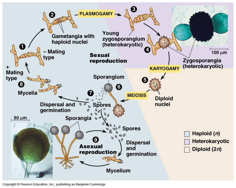
Image 11: Depiction of life cycle of Zygomycota.

Image 12: Pilobolus Day 1- Before experiment setup
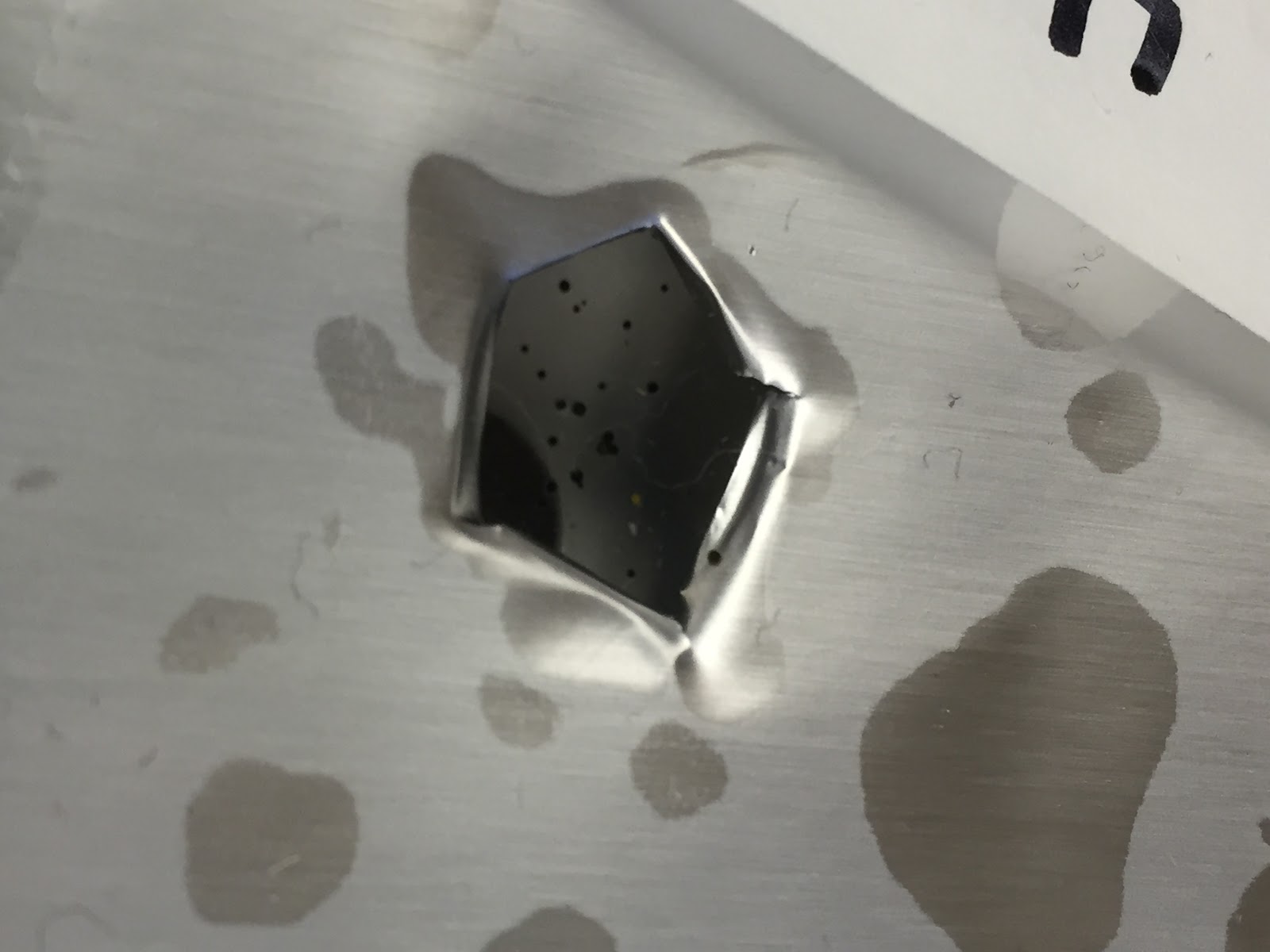
Image 13: The result of the experiment, Pilobolus heads that shot towards the light via phototropism

Image 14: Pilobolus after growth- experiment results
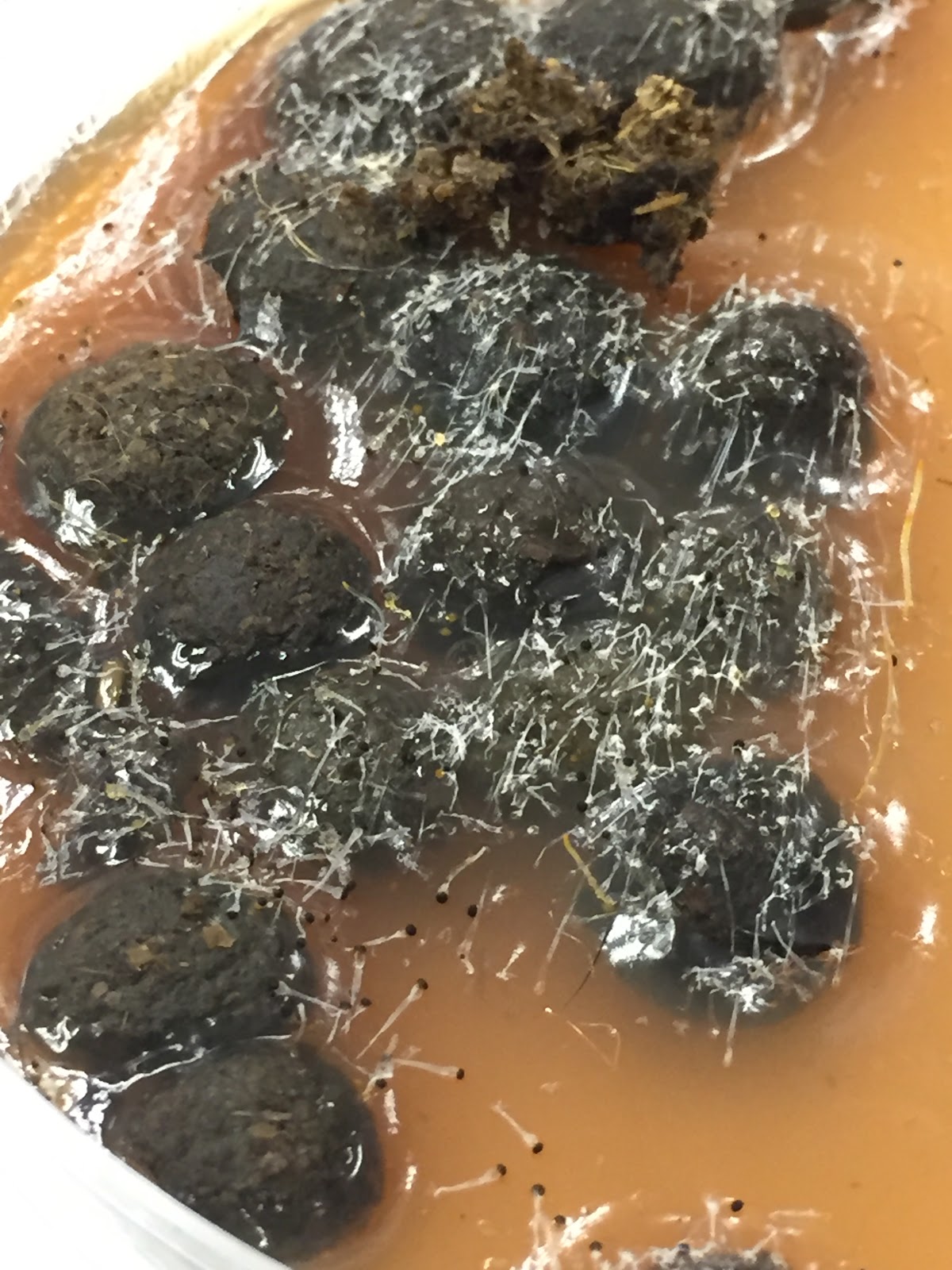
Image 15. Close up of the Pilobolus

Image 16. An even closer look of the Pilobolus- experiment results
Your post is so informative! I had no idea that the Lion's Mane belonged to the Agaricomycetes subdivision, or the genus or kingdom or most of the information about Pilobolus crystallinus.
ReplyDeleteI noticed that your mushrooms didn't bloom that well so I was wondering how often you watered the log, and how much, because my group's log only yielded 4 mushrooms while most other groups had an abundance of them, and we watered ours plenty.
While your mushrooms didn't grow all that much, I assure you, they grew a lot more than my group's (which sadly didn't grow at all and instead decided to rot). It would be interesting to directly compare watering techniques and frequencies of the different groups to determine the best watering mechanisms for mushroom growth and whether it differs between species.
ReplyDelete